Research subpage
Biophotonics
Microscopy, detector engineering, and hyper-dimensional imaging platforms that decode biochemical signatures from fluorescence.
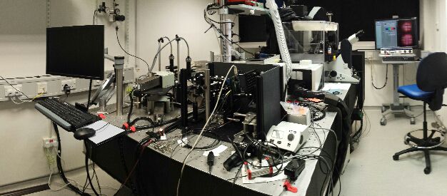
Technology platforms
New horizons in biology and medicine are opened by enabling technologies that allow a deeper understanding of the molecular mechanisms that regulate cellular function and dysfunction. We develop biophysical and biochemical imaging tools that combine emission spectra, fluorescence lifetime, polarization, and engineered instrumentation to decode biochemical signatures from fluorescence.
Our work turns complex assays into practical platforms for specialist and non-specialist laboratories, from biological discovery to drug discovery and diagnostics.
Technology platforms
Live-cell control and fast FLIM
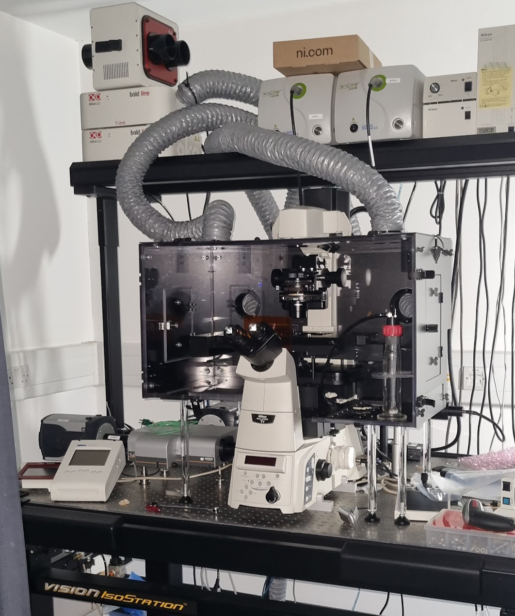
ATLAS.ONE
ATLAS.ONE combines optogenetic perturbation, microfluidics, and fast fluorescence lifetime imaging into a compact platform for controlled live-cell biochemical experiments. It builds on our optogenetic microscope work while remaining distinct from OptoFarm, which is a separate culture and illumination environment.
Project notes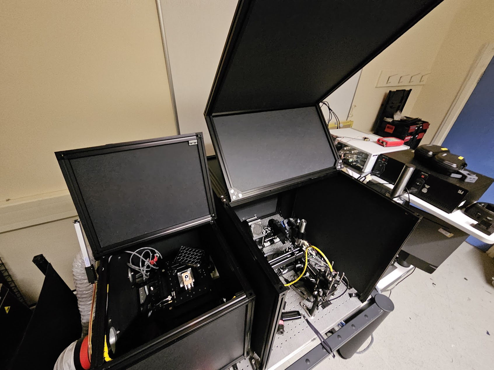
Light-sheet organoid imaging
ATLAS.TWO
ATLAS.TWO brings together the previous ATLAS.TWO concept and OncoLive SPIM direction: a light-sheet platform for imaging organoids and three-dimensional cultures, with optical control of oncogenic events and low-phototoxicity longitudinal observation.
OncoLive archive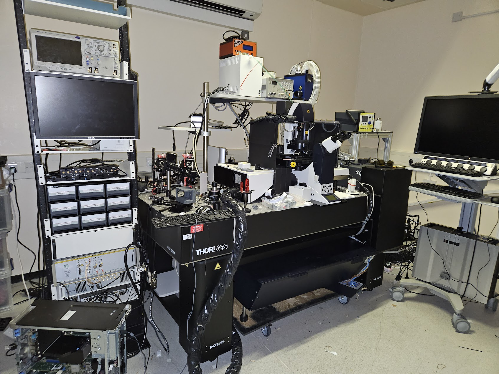
Hyper-dimensional biochemical imaging
ATLAS.THREE
ATLAS.THREE consolidates our HDIM and ELIS work: Leica SP5-based two-photon imaging, multi-colour TCSPC, spectral and polarization-resolved FLIM, and the software-defined detector architecture used to read richer biochemical signatures from living cells and tissues.
Read the HDIM story
Milestones
Milestones
Optogenetics, microfluidics, light-sheet microscopy, and hyper-dimensional FLIM are being organized into ATLAS.ONE, ATLAS.TWO, and ATLAS.THREE.
Spectral, polarization, and lifetime-resolved imaging established the foundations now consolidated in ATLAS.THREE.
A compact optical architecture integrated excitation, emission, and anisotropy spectra for multiplexed fluorescence measurements.
Automation and image cytometry connected molecular readouts to high-content screening workflows.
Solid-state CMOS detection showed that compact, lower-cost lifetime imaging could replace older intensified-camera approaches for many biological applications.
Previous projects
Fast and simple spectral FLIM for biochemical and medical imaging
Collaborative work with David Stoppa at the Fondazione Bruno Kessler in Trento; project outcomes published in 2015 in Optics Express
Abstract | Spectrally resolved fluorescence lifetime imaging microscopy (FLIM) has powerful potential for biochemical and medical imaging applications. However, long acquisition times, low spectral resolution and complexity of spectral FLIM often narrow its use to specialized laboratories. Therefore, we demonstrate here a simple spectral FLIM based on a solid-state detector array providing in-pixel histogramming and delivering faster acquisition, larger dynamic range, and higher spectral elements than state-of-the-art spectral FLIM. We successfully apply this novel microscopy system to biochemical and medical imaging demonstrating that solid-state detectors are a key strategic technology to enable complex assays in biomedical laboratories and the clinic.

Confocal spectropolarimetry
This work was carried out in the laboratories of Prof. Hans Gerritsen, Prof. Clemens Kaminski and Prof. Ashok Venkitaraman; project outcome published in 2011 in Optics Express
Biophysical imaging tools exploit several properties of fluorescence to map cellular biochemistry. However, the engineering of a cost-effective and user-friendly detection system for sensing the diverse properties of fluorescence is a difficult challenge. In this paper, we demonstrated a novel and simple architecture for a spectrograph that permits integrated characterization of excitation, emission and fluorescence anisotropy spectra in a quantitative and efficient manner. This sensing platform achieves excellent versatility of use at comparatively low costs. We demonstrate the novel optical design with example images of plant cells and of mammalian cells expressing fluorescent proteins undergoing energy transfer.
This system is very efficient and, for those interest in fluorescence anisotropy or to analyze spectrally dependent birefringence in materials may find this architecture quite useful. For biological applications, this system would permit to detect homo-FRET, multiplexed over the visible spectrum or to detect changes in fluorescence of environmentally sensitive probes. This is probably one of the most sensitive and simple technique I have developed and the only reason I am currently not using it is that I wanted to allocate my resources on different more ambitious projects. If you are interested to implement this technique, I can share know-how and software (in Matlab).
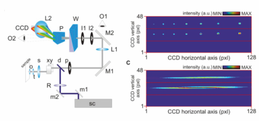
To me, this represents the foundational milestone of multiplexed techniques I am currently developing and I often refer to this system as HDIM-1G (first generation HDIM), albeit the detector I used is an EM-CCD and, therefore, not time-resolved.
Unsupervised FLIM for high throughput imaging of biochemical events
This work was carried out in the laboratory of Prof. Fred Wouters; Click to see paper published in Molecular and Cellular Proteomics (F1000 recommended) in 2007
Proteomics and Cellomics clearly benefit from the molecular insights in cellular biochemical events that can be obtained by advanced quantitative microscopy techniques like fluorescence lifetime imaging microscopy and Foerster resonance energy transfer imaging. The spectroscopic information detected at the molecular level can be combined with cellular morphological estimators, the analysis of cellular localization, and the identification of molecular or cellular subpopulations. This allows the creation of powerful assays to gain a detailed understanding of the molecular mechanisms underlying spatiotemporal cellular responses to chemical and physical stimuli. We demonstrated that the high content offered by these techniques can be combined with the high-throughput levels offered by automation of a fluorescence lifetime imaging microscope setup, capable of unsupervised operation and image analysis. Systems and software dedicated to Image Cytometry for Analysis and Sorting represent important emerging tools for the field of proteomics, interactomics and cellomics.
high content screening by FLIM

Fast wide-field FLIM
This work was carried out in the laboratory of Prof. Fred Wouters in collaboration with CSEM (now spun-off to MESA imaging); project outcomes published in Optics Express in 2005 and in Journal of Biomedical Optics in 2006.
You can also read our patent application or check the wonderful work that PCO have done to make this prototype performing at high standards and commercially available.
Until a few years ago, FD-FLIM systems were limited by the use of expensive technologies and specialized instrumentation, by limited spatial resolution and acquisition throughput and limited capability to resolve heterogeneous systems. We developed methods of analysis and novel technologies to overcome those limitations and to foster the engineering of the new generation of sensing technologies.
Fast and cost-effective system: Solid state technologies for sensing (CMOS and CCD) and sample excitation (LED and laser diodes) were combined in the first cost-effective (~12kEUR + microscope) prototype of an FD imaging system capable of full-field imaging with a single exposure. This technology may replace in the near future the obsolete multi-channel plates used in intensified camera providing fast and efficient FLIM systems. Indeed, PCO is commercializing the first of these cameras (PCO.FLIM) which specs should be good enough for lifetime imaging of biological samples at qualities at least matching those of MCPs.
However, technology is not sufficient to make of wide-field FLIM the technique that we wished: simple, efficient and fast. For this reason, we have developed theoretical frameworks (see Theory section of this website) to optimize it and reach high throughputs and photon-efficiencies (see also papers published in JOSA-A and Biophysical Journal).
 I believe that all these development signed a transition in the community demonstrating for the first time that solid-state technologies developed originally for time-of-flight ranging applications were compatible and mature enough to be redeveloped and applied to lifetime imaging within the life sciences. You can read my thoughts on this topic in this communication to Remote Sensing.